
Assessing Vestibular Disorders
by Judith White, MD, PhD
of Swedish Balance Center
(Presented to Seattle Dizzy Group on 4/9/16)
This presentation gives an overview of vestibular assessment including both common and newer tests and where they fit in the continuum of diagnosis, treatment, and care of patients with vestibular disorders.
Introduction
Dizziness, vertigo, and imbalance can be frustrating for patients to experience and lead to falls. The cause is often readily apparent (for example, Benign Paroxysmal Positional Vertigo or BPPV which can be found with positional testing even without special equipment). Sometimes the cause is more difficult to determine and more detailed evaluation is necessary to determine if there is an inner ear problem, or if attention would be better turned to other causes. Comprehensive vestibular testing is often recommended. Many vestibular tests are available or being developed to understand the complex interaction of eye movements, the brain, balance, and the inner ear.

Evaluation by a vestibular specialist often includes the recording of eye movements using goggles. The inner ear and brain send information to control balance, and keep the eyes on target during head movements. The vestibular system includes the balance portions of the inner ear, central brain and neck connections, and the eye movements and balance reflexes. Examining these eye movements allows the specialist to evaluate the different parts of the vestibular system. Goggle systems allow for recording and examination (usually while patients are briefly in the dark, so that distractions from vision do not interfere with eye movements). Goggle exams are done by many providers, including physical therapists, audiologists and vestibular specialists in neurology, otolaryngology and rehabilitation medicine. Some disorders, such as acute vestibular weakness or BPPV, can be diagnosed using the goggle exam alone.

Comprehensive vestibular testing takes about 90 minutes. It begins with detailed evaluation of eye movements while looking at a large screen TV, and most patients describe this like playing a video game. Next, the ability of the vestibular system and brain to keep the eyes on target during movement is tested using a chair that rotates. The new rotating chairs do not need a surround, since recording accuracy has improved with new technology.
Additional vestibular testing is performed as needed. When comparison of function between the left and right ears needs to be tested, warm and cold air or water may be used to stimulate the inner ears. Tests using clicking sounds (Vestibular Evoked Myogenic Potential or VEMP) are used to check for thinning of the bone around the inner ear, which can cause sound and pressure related vestibular symptoms.
Dizziness
- Most common complaint over age 75
- 8 million annual U.S. visits
- Chronic dizziness (> 2 weeks) affects 16% of self-reported U.S. population.
- 35.4 % of US adults aged 40 and older have vestibular dysfunction (National Health and Nutrition Examination Survey, 2001-2004;Arch Inter Med 169 (10), 938-44, 2009.)
Spiral of Dizziness
- Dizziness increases risk of falling by 8-13 fold
- Limiting activities further increases deconditioning and fall risk (Use it or lose it!)
History
- 70% of dizzy patients can be diagnosed with a careful history (Gufoni, 2005)
- Equilibrium is an unconscious sensation. Few descriptions exist to describe its absence.
- Onset – detailed description of first episode and most recent episode
- Character
- Frequency
- Duration (vertigo and/or milder symptoms)
- Associated hearing loss or tinnitus
- Provoking factors (noise, pressure changes, position change)
Vestibular Labyrinth
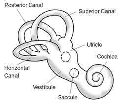
The vestibular labyrinth contains two types of sensors, the semicircular canals and the otolith organs. The semicircular canals sense angular, that is, rotational, movement; specifically, they sense angular acceleration. The otolith organs sense linear motion and orientation with respect to gravity.
There are three semicircular canals, the horizontal, the superior (anterior), and posterior (inferior). Each semicircular canal is sensitive to rotation in its plane. For example, turning the head left and right as if to say “no” stimulates predominantly the horizontal semicircular canal. The planes of the three semicircular canals are nearly perpendicular to one another. Thus, they are able to sense rotation about all axes.
The otolith organs (which contain otoconia) include the utricle and saccule. The utricle senses motion to/fro and left/right and also senses static pitch and roll of the head, i.e. movements like putting one’s chin on the chest or touching ear to shoulder, respectively. The saccule senses up/down motion, to/fro motion, and static pitch of the head.
Note the proximity of the cochlea to the vestibular labyrinth. The vestibular and auditory portions of the inner ear share common blood supply and inner ear fluid. Thus, it is not unusual for disorders that affect the vestibular labyrinth to also affect the cochlea causing hearing loss or tinnitus.
Adapted from Platzer (ed): Pernkopt Anatomy (3rd edition). Urban and Schwarzenberg Inc., Baltimore, 1989.
Otoconia

Otoconia are pebble-like structures composed of crystalized calcium carbonate (CaCO3). The otoconia are constantly being formed and reabsorbed in a process involving the macular supporting cells and surrounding dark cells.
Scanning Electron Micrograph Image of Otoconia from Harada Y (ed): The Vestibular Organs. Amersterdam: Kugler & Guedini, 1988.
Positional Testing


Otoconia (also known as particles or canaliths) may migrate into the semicircular canals, trigger nerve receptors, and cause positional vertigo.
- Lateral supine head turns and Dix-Hallpike positioning (to provoke/evaluate nystagmus indicative of positional vertigo)
- Latency
- Duration
- Fatigue
- Habituation
Benign Paroxysmal Positional Vertigo (BPPV)
- Vertigo of sudden onset and brief duration provoked by changes in head position
- Lying down
- Rolling over in bed
- Bending over
- Looking up
- Washing hair in shower
- Dentist or beauty parlor
Incidence/Prevalence of BPPV
- Most common referred diagnosis in tertiary centers
- 9% of randomly selected community dwelling elderly (Oghalai, J.S. et al 2000)
- Incidence increases 38% with each decade of life (Froehling 1991)
Peripheral Nystagmus
- Jerk nystagmus (slow and fast phase)
- Direction fixed
- Generally beats away from affected ear
- Worse in the direction of gaze that is towards the fast phase
National Guidelines for BPPV
- American Academy of Otolaryngology and American Academy of Neurology
- Perform the Dix-Hallpike maneuver for diagnosis
- Recommend not performing radiological imaging
- Recommend against use of vestibular suppressant medication
- Perform repositioning maneuver and/or vestibular rehabilitation referral
- Evaluate treatment failures for underlying vestibular or CNS disorders
Canalith Repositioning Procedure
(otherwise known as Particle Repositioning Maneuver)

BPPV can affect any of the three canals in each ear, and examination of the eye movements (nystagmus) during positioning testing can help determine which canals are affected. In turn, maneuvers can be performed using video goggles for guidance to maximize the success of the repositioning.
BPPV Recurrence
- Recurrence is common (15%/yr)
- Otoconial adherence or mineralization/ demineralization abnormalities may contribute to recurrence
- Home canalith repositioning is effective (Radke, 2004)
Vestibulo-Ocular Reflex

Posturography

- Postural control is a complex interplay of visual, proprioceptive and vestibular input
- Posturography tests static postural control in a series of conditions designed to emphasize or minimize each of these inputs
Caloric Tests
- Only used to compare the inner ear function on one side to that on the other side
- Used less often due to newer technologies
- Subject to technical artifact
- May be necessary before procedures to verify normal vestibular function in the good ear
Acute Vestibular Syndrome
- Rapid unilateral injury to either peripheral or central vestibular structures produces prolonged vertigo (days to weeks)
- Severe vertigo, nausea and vomiting, spontaneous nystagmus and postural instability
- Usually peripheral and can be associated with hearing loss (labyrinthitis)
- Timely diagnosis improves recovery (ASAP prednisone 1mg/kg/day for ten days and taper 10 mg/day – Strupp M., 2004 NEJM)
- Avoid vestibular suppressants after 3 days and begin vestibular rehabilitation to hasten compensation.
- Compensation from the brain and good ear begin after three days
- Vestibular suppressants delay compensation
- Compensation can be measured by looking at eye movements while moving the head
Vestibular Evoked Myogenic Potential (VEMP)

Clinical Applications
- The Cervical-VEMP is a test of saccular function
- Inferior vestibular nerve response is assessed
- Complements vestibular test procedures that assess lateral semicircular canal/superior division of the vestibular nerve.
- Asymmetric low thresholds are most predictive of SSCD
Superior Semicircular Canal Dehiscence (SSCD)
- First described by Minor et al 1998
- Sound or pressure induced eye movements
- Defect in the bone overlying the SCC forms an abnormal connection between the vestibular system and intracranial compartment.
Common Presenting Symptoms/Complaints
- Pressure Induced Disequilibrium
- Elevators
- Scuba Diver
- Sneezing
- Flying
- Straining
- Lifting Weights
- Sound induced disequilibrium
- Loud noises cause imbalance or frank vertigo
- Organ music
- Fire alarm
- School bells (teachers)
- Loud telephone or music
- Audiologist (demonstrating audiologic screening on self)
- Loud noises cause imbalance or frank vertigo
- Hearing Loss
- Muffled, blocked or echo in hearing
- Usually low frequency
- Typically reduced hearing for sounds presented thru the air, but unusually sensitive hearing for sounds presented via vibration (bone conduction)
- Patient may hear a vibrating tuning fork applied to their ankle in their ear (conducts vibration thru skeleton)
Eye Movements Align with the Abnormal Canal


- Tonic upwards and ipsilateral intorsion is seen with ampullofugal endolymph flow – activation (loud noise, positive middle ear pressure)
- Tonic downwards and ipsilateral extorsion is seen with ampullopetal endolymph flow – inhibition (Valsalva against a closed glottis, jugular compression, raised intracranial pressure)
Vestibular Assessment Summary
- Vestibular tests help evaluate the location of any problems in the inner ear
- Vestibular tests are also helpful in evaluating other central nervous system disorders affecting eye movement and balance
 Judith White, MD, PhD
of Swedish Balance Center
Dr. Judith White listens carefully and focuses on the details of dizziness, vertigo and balance problems, in order to provide efficient and accurate diagnosis and convenient evidence-based treatment. The Swedish Balance Center uses a team approach, with specialists in ear, balance and rehabilitation working together, with the full resources of the Swedish Neuroscience Institute. Preventing falls and their potentially life-altering consequences is the priority of our work, consistent with the Centers for Disease Control and Prevention national priorities.
More information about Dr. White
*******
Presentation information is not meant to be taken as medical advice.
Presentations posted online may include discussion notes, links, images, and other information added by Seattle Dizzy Group.
*******
Post updated May 2, 2016
© Copyright 2016, Seattle Dizzy Group. All rights reserved.


[…] 2016, we had guest speakers on the topics of Reducing Inflammation, Vestibular Testing & Assessment, and Vision […]
By: Stronger Together in 2017 | Seattle Dizzy Group on January 31, 2017
at 10:43 pm
[…] Vestibular Testing […]
By: Treating Dizziness and Imbalance with Physical Therapy | Seattle Dizzy Group on October 31, 2017
at 11:57 pm